| Line 26: | Line 26: | ||
The second assumption is that the energy loss from absorbed photons is significant. Each photon that travels through the material has a probability of being "absorbed" for each increment of distance it travels. Being "absorbed" means that the photon is scattered, changing its direction and wavelength enough for the detector to miss that photon. The scattered photons dissipate into the surrounds in the form of heat. These two assumptions imply that photons emitted from the X-ray source either gets to the detector directly opposite the pinhole, or it doesn't. | The second assumption is that the energy loss from absorbed photons is significant. Each photon that travels through the material has a probability of being "absorbed" for each increment of distance it travels. Being "absorbed" means that the photon is scattered, changing its direction and wavelength enough for the detector to miss that photon. The scattered photons dissipate into the surrounds in the form of heat. These two assumptions imply that photons emitted from the X-ray source either gets to the detector directly opposite the pinhole, or it doesn't. | ||
| + | [[Image:collimator.jpg|200px|thumb|left|Figure 4: Photon Path and Attenuation]] | ||
| + | ---- | ||
References:<br /> | References:<br /> | ||
Revision as of 21:11, 15 December 2014
Computed Tomography (CT)
A slecture by ECE student Sahil Sanghani
Partly based on the ECE 637 material of Professor Bouman.
Introduction
Computed Tomography (CT) is a X-ray based method of 3D imaging. Other 3D imaging techniques are Magnetic Resonance Imaging (MRI) and Positron Emission Tomography (PET). Both CT and PET use tomographic reconstruction to create their 3D representations. Tomography has a variety of uses in the medical field, remote nondestructive sensing, RADAR, and SAR.
CT Scanner Structure
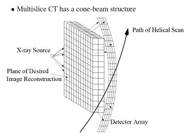
Figure 1 shows a typical CT scanner used for medical imaging. The CT scanner is composed to two main parts: the bed and the scanner. The bed horizontally translates through the scanner, which houses the rotating X-ray source and detector. Figure 2 shows a CT scanner without the cover. The X-ray source, detector and travel have been highlighted. As the bed translates horizontally through the scanner, the scanner collects multiple data scans and processes them. Because the scanner is collecting as the patient moves through it, the path of the scan is actually helical, as shown in Figure 3.
Photon Attenuation
There are a couple assumptions and approximations that are used in the CT scanning and recording technology. The first approximation is that the X-ray source emits photons that supposedly travel in a straight line. This can be achieved with a pinhole collimator as shown in Figure 4. Just like how a lens focuses light, the pinhole collimator focuses X-rays. Typically a pinhole isn't used because the diffraction effects cause constructive and destructive interference. However, since the wavelength of X-rays is so small, the diffraction artifacts don't affect the pinhole collimator. Thus we can assume that the X-rays that come through the pinhole propagate in a straight line.
The second assumption is that the energy loss from absorbed photons is significant. Each photon that travels through the material has a probability of being "absorbed" for each increment of distance it travels. Being "absorbed" means that the photon is scattered, changing its direction and wavelength enough for the detector to miss that photon. The scattered photons dissipate into the surrounds in the form of heat. These two assumptions imply that photons emitted from the X-ray source either gets to the detector directly opposite the pinhole, or it doesn't.
References:
[1] C. A. Bouman. ECE 637. Class Lecture. Digital Image Processing I. Faculty of Electrical Engineering, Purdue University. Spring 2013.
[2] Fung, B. 2012. The Atlantic. December 16, 2014. from: http://www.theatlantic.com/health/archive/2012/09/how-to-build-your-own-ct-scanner/262066/
Note: Photo Edited by Sahil Sanghani


