| Line 16: | Line 16: | ||
---- | ---- | ||
=CT Scanner Structure= | =CT Scanner Structure= | ||
| − | + | [[Image:CT_scanner.jpg|200px|thumb|left|Fig 1: A CT Scanner]] | |
| + | [[Image:CT_Internals_labeled_scaled.jpg|300px|thumb|right|Fig 2: CT Scanner Internals]] | ||
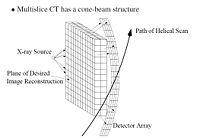
Figure 1 shows a typical CT scanner used for medical imaging. The CT scanner is composed to two main parts: the bed and the scanner. The bed horizontally translates through the scanner, which houses the rotating X-ray source and detector. Figure 2 shows a CT scanner without the cover. The X-ray source, detector and travel have been highlighted. As the bed translates horizontally through the scanner, the scanner collects multiple data scans and processes them. Because the scanner is collecting as the patient moves through it, the path of the scan is actually helical, as shown in Figure 3. | Figure 1 shows a typical CT scanner used for medical imaging. The CT scanner is composed to two main parts: the bed and the scanner. The bed horizontally translates through the scanner, which houses the rotating X-ray source and detector. Figure 2 shows a CT scanner without the cover. The X-ray source, detector and travel have been highlighted. As the bed translates horizontally through the scanner, the scanner collects multiple data scans and processes them. Because the scanner is collecting as the patient moves through it, the path of the scan is actually helical, as shown in Figure 3. | ||
| − | + | [[Image:HelicalSlice.jpg|200px|center|Fig 3: Multislice Helical Scan CT|]] | |
---- | ---- | ||
References:<br /> | References:<br /> | ||
| Line 27: | Line 28: | ||
| − | [[ HonorsContractECE438Fall14|Back to | + | [[ HonorsContractECE438Fall14|Back to Honors Contract Main Page]] |
Revision as of 19:34, 15 December 2014
Computed Tomography (CT)
A slecture by ECE student Sahil Sanghani
Partly based on the ECE 637 material of Professor Bouman.
Introduction
Computed Tomography (CT) is a X-ray based method of 3D imaging. Other 3D imaging techniques are Magnetic Resonance Imaging (MRI) and Positron Emission Tomography (PET). Both CT and PET use tomographic reconstruction to create their 3D representations. Tomography has a variety of uses in the medical field, remote nondestructive sensing, RADAR, and SAR.
CT Scanner Structure
Figure 1 shows a typical CT scanner used for medical imaging. The CT scanner is composed to two main parts: the bed and the scanner. The bed horizontally translates through the scanner, which houses the rotating X-ray source and detector. Figure 2 shows a CT scanner without the cover. The X-ray source, detector and travel have been highlighted. As the bed translates horizontally through the scanner, the scanner collects multiple data scans and processes them. Because the scanner is collecting as the patient moves through it, the path of the scan is actually helical, as shown in Figure 3.
References:
[1] C. A. Bouman. ECE 637. Class Lecture. Digital Image Processing I. Faculty of Electrical Engineering, Purdue University. Spring 2013.
[2] Fung, B. 2012. The Atlantic. December 16, 2014. from: http://www.theatlantic.com/health/archive/2012/09/how-to-build-your-own-ct-scanner/262066/
Note: Photo Edited by Sahil Sanghani


